.jpg)
Case Study 1
Veterinary Examination
This study describes a visit to a veterinary specialist with a cat suffering from small cell lymphoma. The visit included both X-ray and ultrasound examinations.
Case Study 2
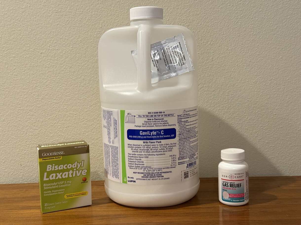
EGD and Colonoscopy
This study describes a visit to the hospital for a routine esophagogastroduodenoscopy (EGD) and colonoscopy.
.jpg)
.jpg)