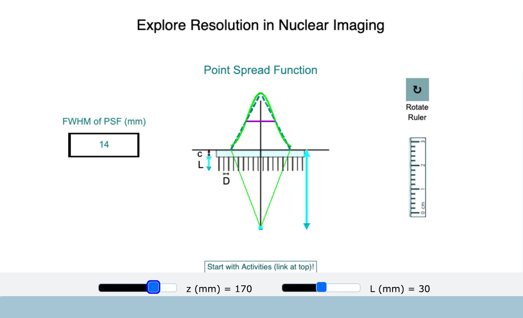
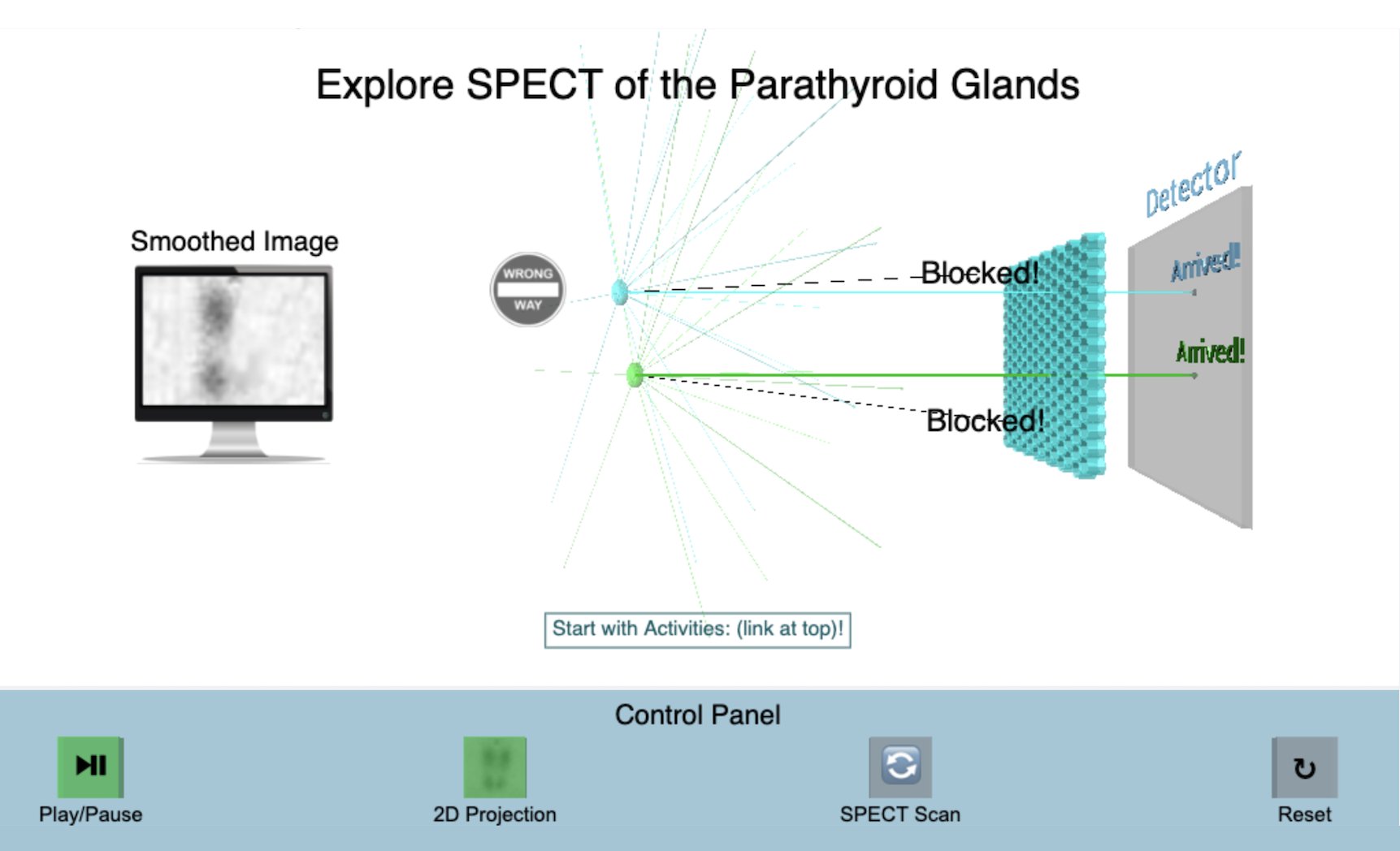
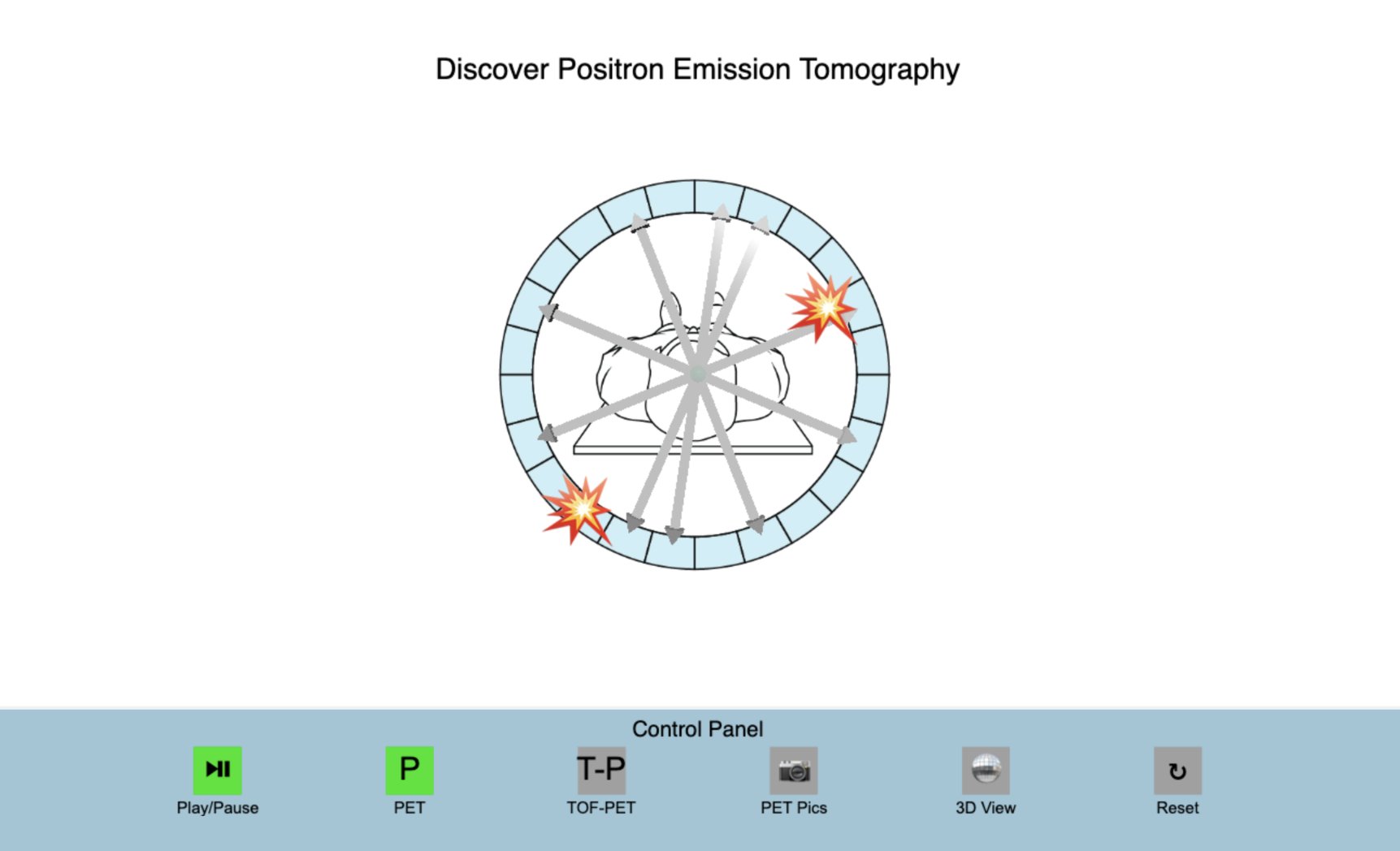
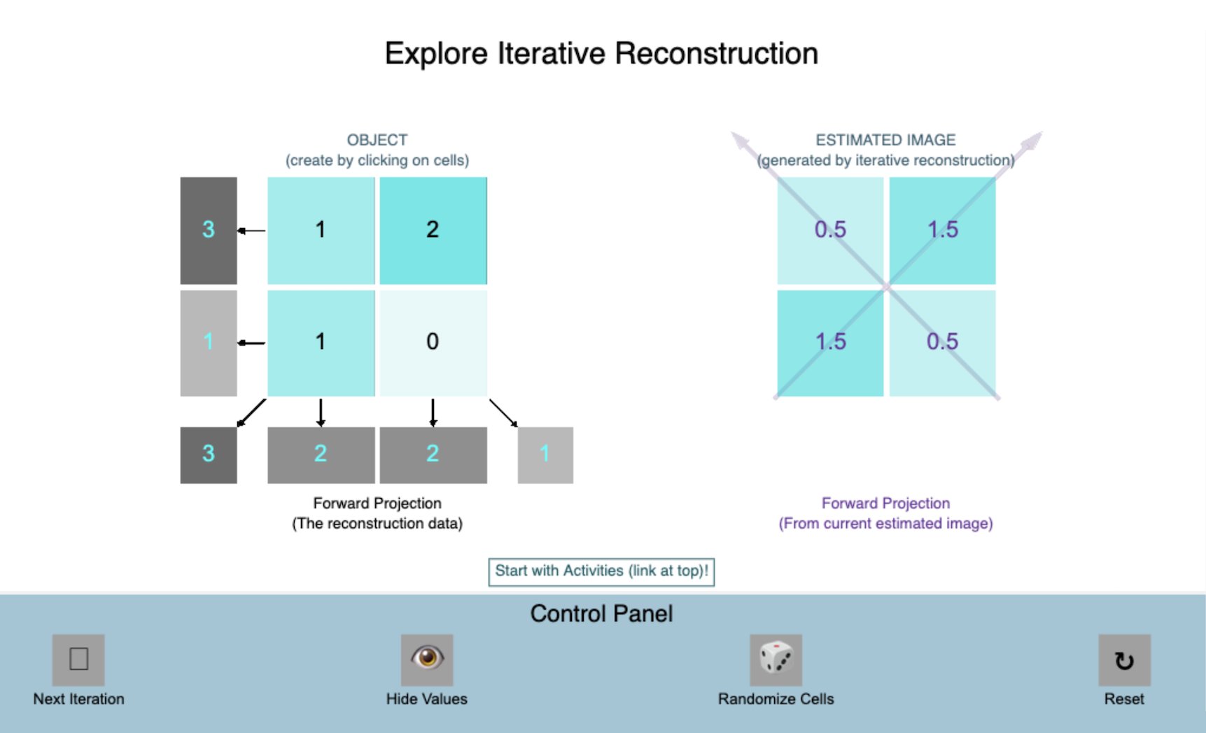
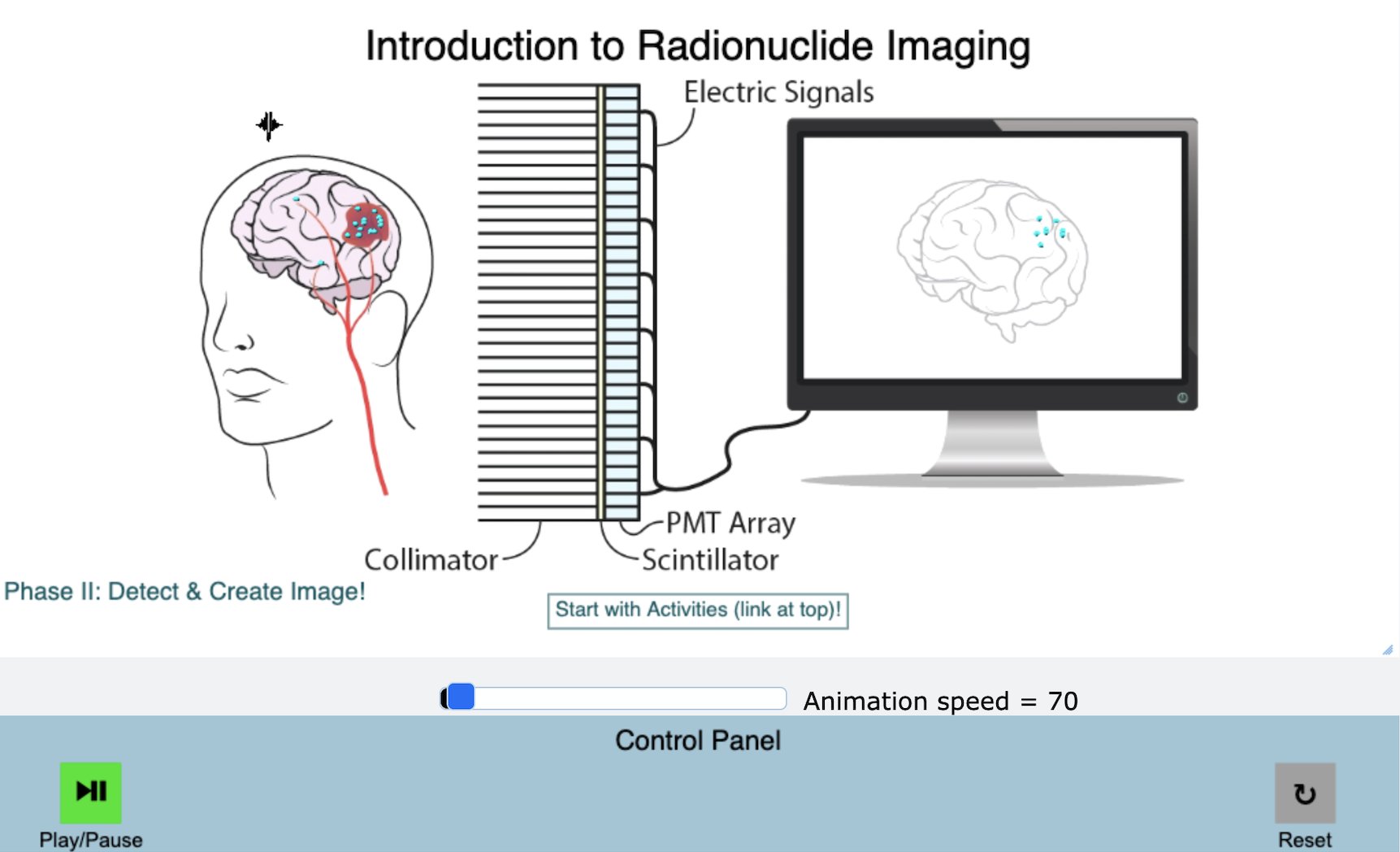
Explore the fundamentals of radionuclide imaging, including (a) labeling a patient with a radiopharmaceutical (i.e., a pharmaceutical tagged with a radioactive element), (b) delivery of the pharmaceutical to a site of interest (e.g., a tumor) in the body, (c) detection of gamma rays emitted during radioactive decay, and (d) creation of an image from detected gamma ray counts.
Explore the fundamentals of radioactivity, including (a) the existence of isotopes, (b) mechanisms leading to nuclear instability (radioactivity), (c) the importance of gamma ray photons in image creation, (d) direct gamma emission by technetium-99m, (e) indirect gamma creation following positron emission by fluorine-18, and (f) direct gamma and beta emission by iodine-131.
Explore the fundamentals of resolution in radionuclide imaging, including the roles of (a) source depth, (b) collimator diameter, and (c) collimator length
Explore the fundamentals of projection and tomographic nuclear imaging, including (a) generation of a projection (view) on a detector, (b) roles of collimation in projection creation and signal loss, (c) limitations of projection-based imaging (e.g., loss of depth information), (d) complementary anatomical information provided by projections collected at different viewing angles, and (e) generation of depth-preserving slices and 3D images using emission computed tomography (ECT)
Explore the fundamentals of positron emission tomography (PET), including (a) generation of two anti-parallel gamma rays following positron emission from an unstable nucleus, (b) absence of a collimator, (c) line of response (LOR) creation along the line connecting the two detectors impacted by the anti-parallel gamma rays, and (d) the role of time-of-flight (TOF) technology in improving signal-to-noise and definition of small lesions in images (as well as reducing injected dose and scan times)
Explore the mathematical basics of image reconstruction in ECT, including (a) generation of projection data for an object, (b) iterative reconstruction of the activity distribution in the object, and (c) comparison of estimated and measured projection data and use of the difference to update and improve the activity estimate.