Introductory Biomedical Imaging

Magnetic Resonance Imaging Simulations

Magnetic Resonance Imaging Simulations

Magnetic resonance imaging (MRI) is an imaging variant of nuclear magnetic resonance (NMR). MRI is renowned for its high-resolution, exquisite soft-tissue contrast, ability to monitor anatomy and function, and safety. MRI uses an unusual localization strategy to visualize protons in water in living tissue: signals arising from protons in different regions in the body are distinguished, and their distinct points of origin are identified, based on frequency. This approach, which is known as frequency encoding, is implemented by placing protons in a spatially varying magnetic field, which creates signals with frequences that are determined by their location. Like other approaches, image contrast in MRI is based on differences in signal amplitudes. One parameter that affects signal amplitude is proton density, which can be used as a source of contrast. However, soft tissues tend to have similar proton densities; thus, “density-weighted” images are relatively low in contrast. MRI’s strength for soft-tissue imaging arises because the MRI signal amplitude can be made to depend on other parameters, such as the local environment, that generate much better contrast. Weaknesses of MRI include relatively slow data acquisition, high equipment cost, and incompatibility with metal implants. MRI is notable for providing both anatomic and functional information.

The following simulations explore nuclear magnetism and how time-varying magnetic fields can be used to create structural and functional images. The simulations are listed in the recommended order of performance. Each simulation includes a home page from which the simulation can be run, together with links to simulation-specific Information, Background, and Activity documents.


...

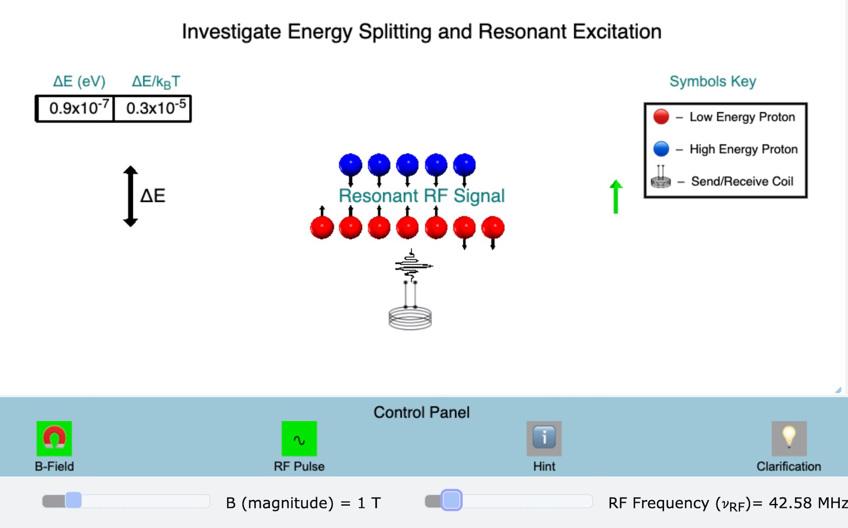
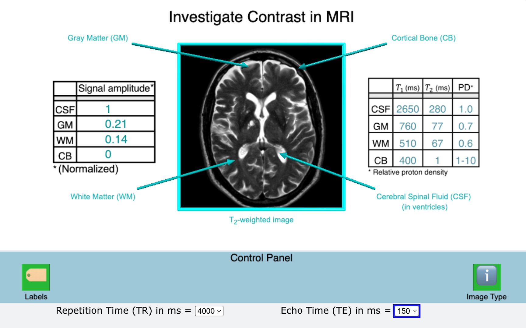
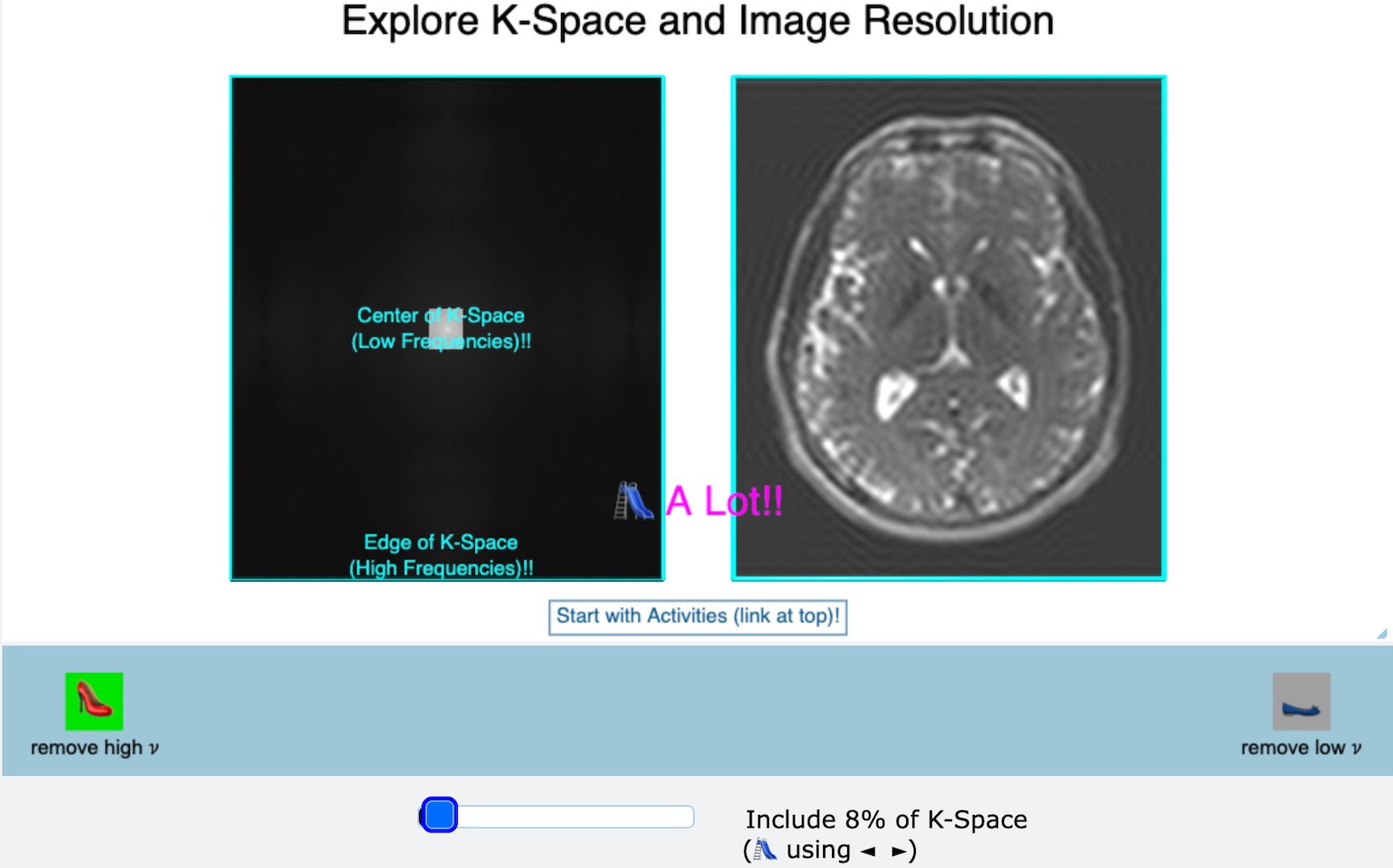
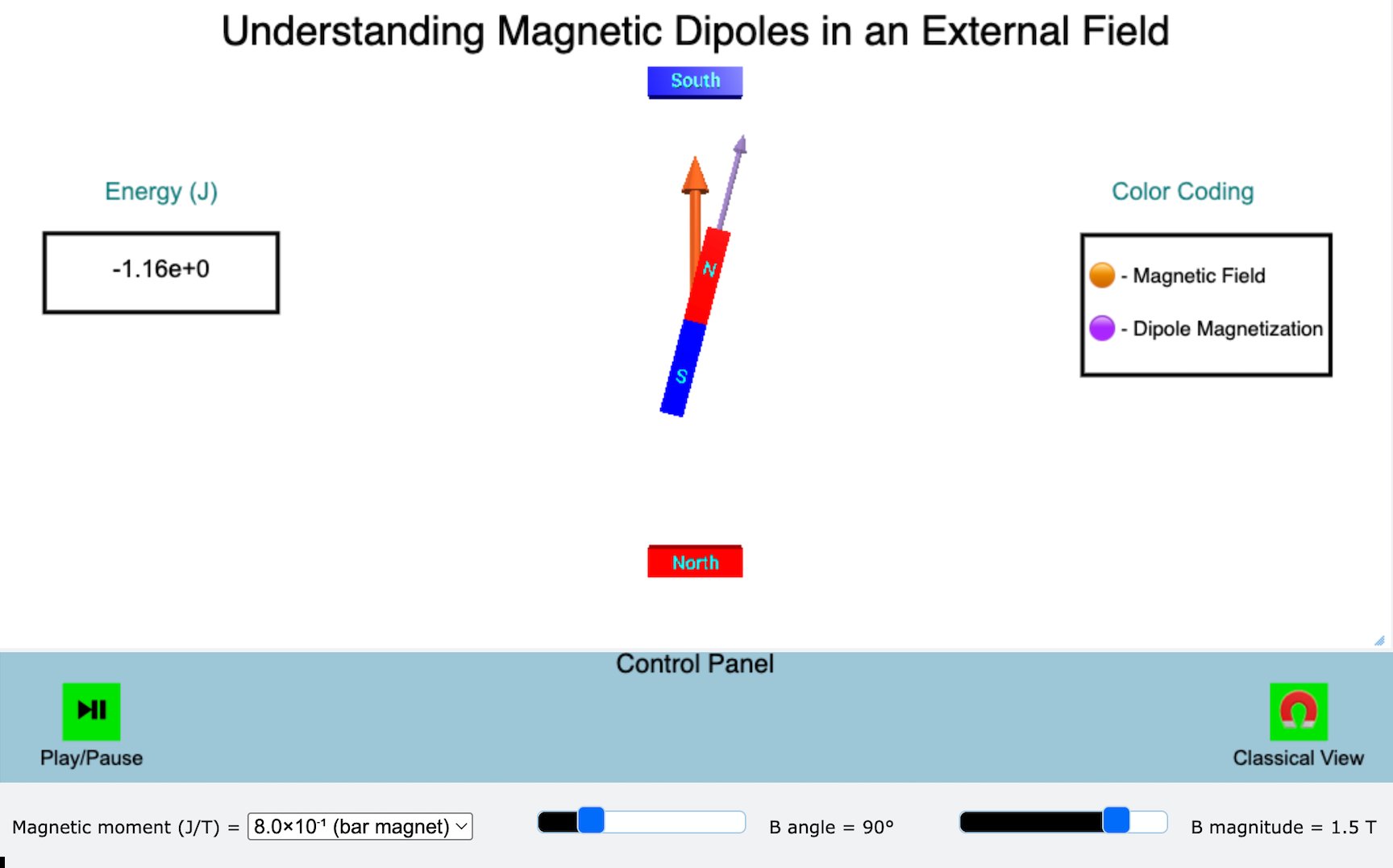
Nuclear Magnetic Dipoles

Explore the fundamentals of nuclear magnetism and interactions between magnetic dipole moments and magnetic fields, including (1) existence of intrinsic nuclear magnetism and magnetic moments, (b) energy of interaction between a magnetic moment and an external magnetic field, (c) energy minimization and maximization when the magnetic moment and field are parallel and anti-parallel, respectively, and (d) the tendency for a magnetic moment to align parallel to an external magnetic field

...

Resonant Excitation

Explore the fundamentals of energy quantization and resonance excitation in MRI, including (a) unbiased alignment of magnetic moments and energy-level “degeneracy” (i.e., the presence of only one energy level) in the absence of an external magnetic field, (b) slightly biased alignment and energy level splitting (into two distinct levels) in the presence of an external magnetic field, and (c) requirement for resonant excitation to generate an MRI signal

...

Field Gradients and 1D Imaging

Explore the fundamentals of field gradients, frequency encoding, and 1D projection imaging in MRI, including (a) gradient magnetic fields with a magnitude that varies linearly with position, (b) Associated resonance excitation and emission frequencies that also vary linearly with position, and (c) projection imaging with just one field gradient

...

2D and 3d Imaging

Explore the fundamentals of 2D/3D Imaging in MRI, including (a) slice selection using a gradient and an RF pulse with a defined center frequency and frequency range (width), (b) roles of pulse width and gradient strength in determining slice width along , (c) the role of pulse center frequency in determining slice location along , (d) localization within the slice using two additional field gradients along and , and (e) phase and frequency encoding

...

Classical Description of Signal Generation and Decay

Explore the fundamentals of classical signal generation and decay in MRI, including (a) an initial equilibrium configuration with net magnetization along the external field, (b) creation of a non- equilibrium configuration via energy input from an on-resonance RF pulse, (c) decay of the MRI signal over a tissue-dependent time-scale T2, and (d) recovery of equilibrium over a tissue- dependent time-scale T1.

...

Contrast in MRI

Explore the fundamentals of contrast generation in MRI, including (a) the role played by pulse sequences, (b) generation of relaxation time– and proton density–weighted contrast with appropriate choices of signal measurement time and experiment repetition time for a pulse sequence, and (c) differences in tissue brightness that result from different kinds of contrast weighting

...

K-space, Resolution, and Image Reconstruction

Explore the fundamentals of K-space data and its relationship to image resolution.