Introductory Biomedical Imaging

X-Ray Imaging Simulations

X-Ray Imaging Simulations

Conventional radiography and computed tomography (CT) are closely related techniques. Images are generated by detecting X-rays that traverse a patient positioned between a source and a detector. X-ray transmission is high for radiolucent regions of the body, such as the lungs, which lack X-ray absorbing structures. In contrast, X-ray transmission is low for radiopaque regions, such as bone. In X-ray images, radiolucent areas appear black, and radiopaque areas appear white. In conventional radiography, each image point reflects summed X-ray attenuation along an associated straight-line trajectory between the source and detector. The resulting “projection” images lack depth information and suffer from structural superposition. In contrast, in CT, images are created by collecting projections at many viewing angles and using the different views to reconstruct high-resolution, depth-preserving two-dimensional slices and three-dimensional images of anatomical structures, including soft tissue. Conventional radiography has many uses, including detecting broken bones, cavities, and various cancers. CT also has many uses, including rapid emergency assessments, cancer diagnosis and treatment, and in conjunction with radionuclide imaging to create hybrid structural/functional images. Both conventional radiography and CT are notable for providing anatomic (i.e., structural) information.

The following simulations explore the foundations of X-ray imaging and how X-rays are used to create both planar and tomographic images. The simulations are listed in the recommended order of performance. Each simulation includes a home page from which the simulation can be run, together with links to simulation-specific Information, Background, and Activity documents.


...

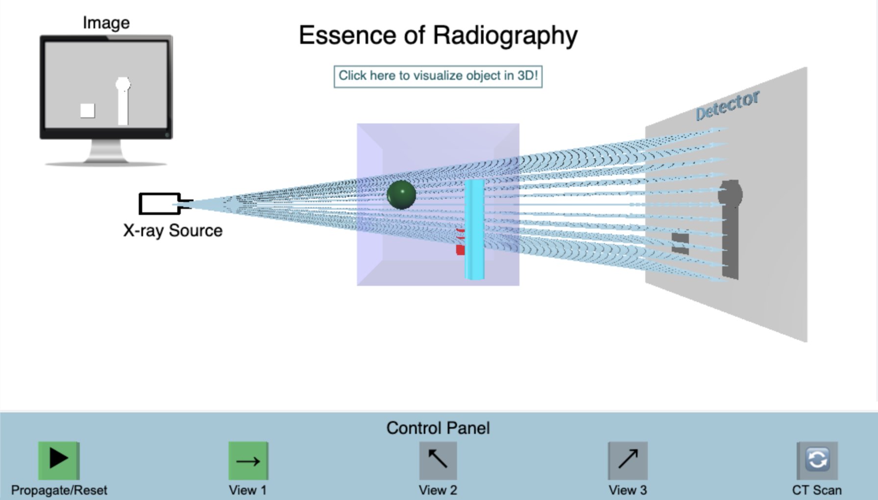
Essence of Radiography

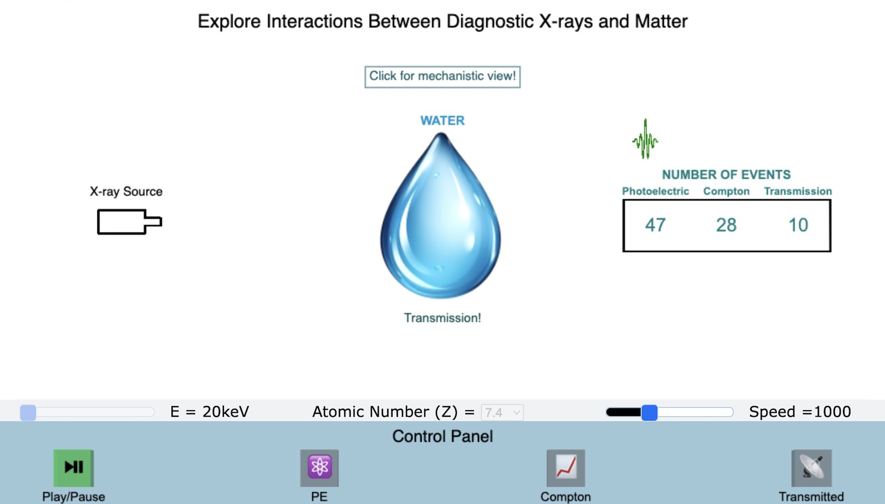
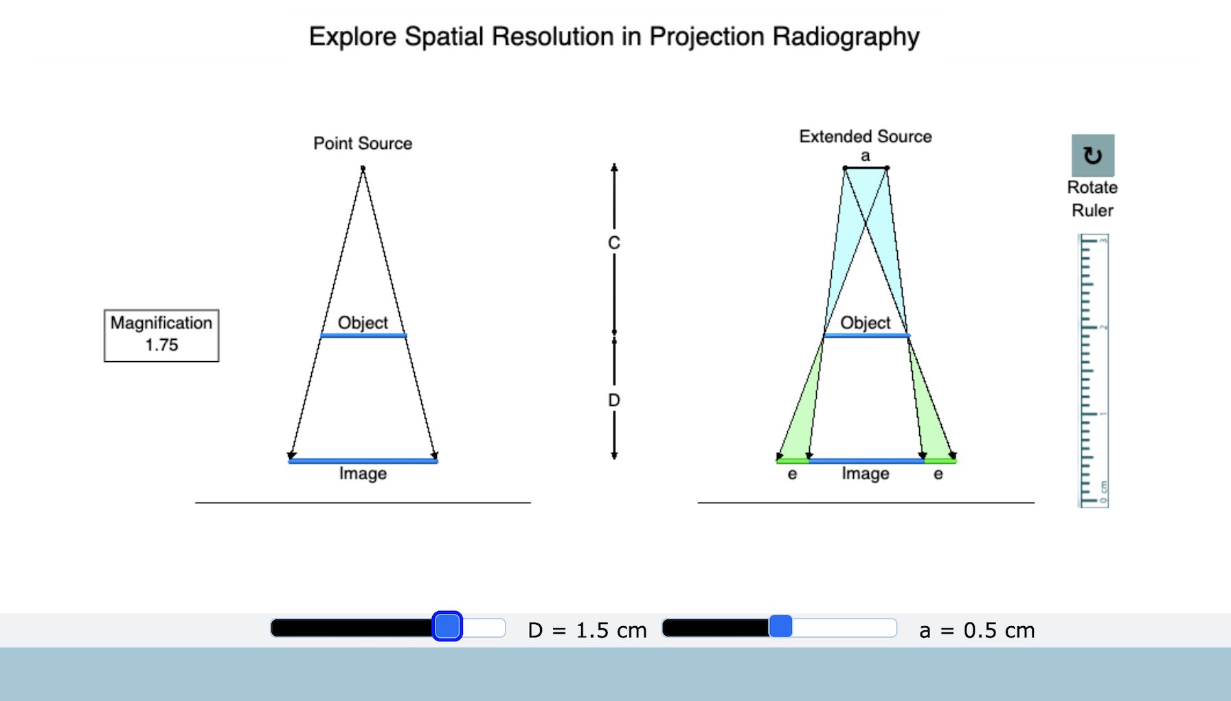
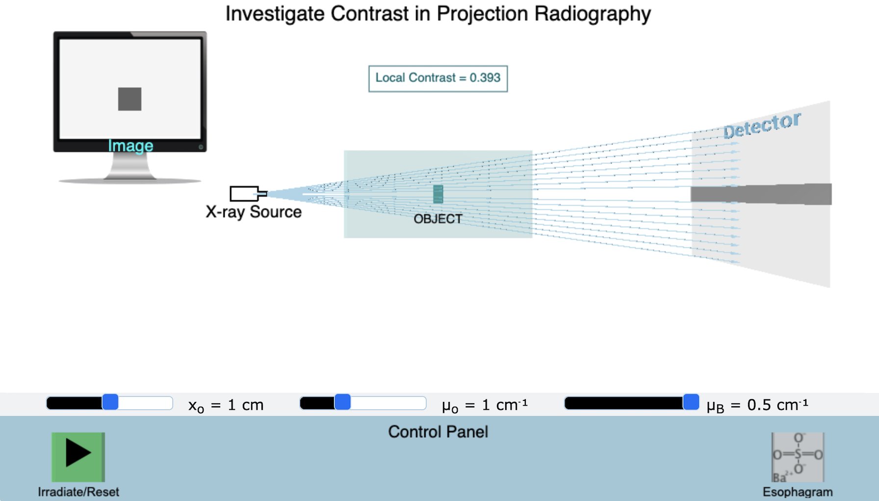
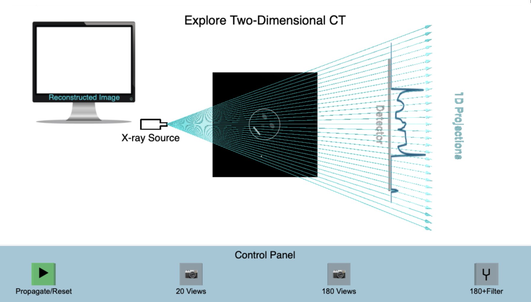
Explore the fundamentals of X-ray-based imaging, including (a) generation and detection of X- rays, (b) creation of a projection (view) on a detector, (c) limitations of projection-based imaging (e.g., loss of depth information), (d) complementary anatomical information provided by projections collected at different viewing angles, and (e) generation of depth-preserving slices using computed tomography (CT).

...

Interactions Between Diagnostic X-Rays and Matter

Explore the physical mechanisms that govern the creation of diagnostic images using X-rays, including (a) absorption via the Photoelectric Effect, (b) scattering via the Compton Effect, (c) transmission of non-interacting X-rays, and (d) effects of X-ray energy and atomic number on interaction probabilities and transmission.

...

Resolution in Diagnostic Radiography

Explore the fundamentals of resolution in diagnostic radiography, including (a) the roles of source size and object-to-detector separation in determining resolution, and (c) the relationship between resolution and magnification.

...

Contrast in Diagnostic Radiography

Explore the fundamentals of contrast in diagnostic radiography, including the effects of (a) object thickness, (b) mismatch in attenuation coefficients, (c) photon energy, and (d) contrast media (the last as exemplified in an esophagram).

...

Computed Tomogaphy

Explore the fundamentals of image reconstruction in CT, including (a) generation of one- dimensional projection data for a two-dimensional object, (b) creation of a reconstruction of the object via inverse radon transformation, and (c) the effects of the number of projections and of filtering on reconstruction quality.

...

Image Reconstruction via Simple Back Projection

Explore the fundamentals of image reconstruction in CT, including (a) generation of forward projection data for an object, and (b) generation of a blurred reconstruction of the object via simple back projection.